Thursday, June 23, 2016

Body-Mass Index in 2.3 Million Adolescents and Cardiovascular Death in Adulthood

AIM
- To investigate the association between body mass index (BMI) in late adolescence and death from cardiovascular disease in adulthood.

METHOD
-BMI data from 1967 through 2010 Israeli adolescents (mean age 17.3±0.4 years) were grouped according to age- and sex- specific percentiles from the US CDC.
[Underweight: <5th percentile
Normal weight: 5th to 24th percentile (Reference group), 25th to 49th percentile, 50th to 74th percentile, 75th to 85th percentile
Overweight: 85th to 94th percentile
Obese: ≥ 95th percentile]
- Total number of study sample: 2,298,130 participants
- Primary outcomes: number of deaths attributed to coronary heart disease, stroke, sudden death from unknown cause or a combination of all three categories (total cardiovascular causes) by mid-2011.
- Statistical analysis: Cox proportional-hazards models; imputed 1.4% of missing values (country of origin, education and socioeconomic status) using multiple imputation algorithm; applied linear and quadratic terms using the midpoint of each BMI percentile group or the median of each absolute BMI group; spline models were fit to estimate the BMI value associated with minimum cardiovascular mortality and the lowest BMI associated with significantly increased mortality.

RESULTS
- During 42,297,007 person-years of follow-up, 2918 of 32,127 deaths (9.1%) were from cardiovascular causes, including 1497 from coronary heart disease, 528 from stroke, and 893 from sudden death.
- The mean ages at the time of death were 47.4 years for coronary heart disease, 46.0 years for stroke, and 41.3 years for sudden death
- The rates of death per person-year were generally lowest in the group that had BMI values during adolescence in the 25th to 49th percentiles, with higher rates observed among those below the 5th percentile.
- A graded increase in the risk of death from cardiovascular causes and all causes that started among participants in the group that was in the 50th to 74th percentiles of BMI (i.e., within the accepted normal range).

Analysis based on percentile grouping
Hazard ratio after multivariable adjustment* (Reference group: 5th to 24th percentile):
- Overweight (85th to 94th percentiles): 3.0 (95% CI, 2.5-3.7) for death from coronary heart disease, 1.8 (95% CI, 1.3-2.5) for death from stroke, 1.5 (95% CI, 1.1-1.9) for sudden death, and 2.2 (95% CI, 1.9-2.6) for death from total cardiovascular causes
-Obese group (≥95th percentile for BMI): 4.9 (95% CI, 3.9-6.1) for death from coronary heart disease, 2.6 (95% CI, 1.7-4.1) for death from stroke, 2.1 (95% CI, 1.5-2.9) for sudden death and 3.5 (9.5% CI, 2.9-4.1) for death from total cardiovascular causes
*(adjustment for sex, age, birth year, sociodemographic characteristics, and height)

Analysis based on year of follow-up
- Hazard ratio from death from cardiovascular disease increased from 2.0 (95% CI 1.1 to 3.9) during follow-up for 0 to 10 years to 4.1 (95% CI 3.1 to 5.4) during follow-up for for 30 to 40 years
- The risk was persistently high for death from coronary heart disease across the entire period of follow-up.

Analysis of absolute values of BMI
- Participants with a BMI ranging from 20.0 to 22.4 had a higher risk of death from coronary heart disease than did those with a BMI ranging from 17.5 to 19.9 (hazard ratio, 1.2; 95% CI, 1.1-1.4)
- The risks of death from stroke, sudden death, and total cardiovascular causes (along with noncardiovascular causes and all causes) were also elevated starting with a BMI of 22.5 and increased more steeply among the extremely obese for cardiovascular-specific death
On multivariable-adjusted spline models,
-The minimum risks of death from stroke, sudden death, and cardiovascular causes were among participants who had BMI values of 19.8, 19.3, and 18.3, respectively, whereas the association with death from coronary artery disease was graded across the full range of BMI-percentile groups
- Participants had a significantly elevated risk of death from total cardiovascular causes starting at BMI values above 20.3.

Analysis by z-scores of BMI
- After multivariable adjustment, the hazard ratios for death from coronary heart disease were 1.54 (95% CI, 1.46 to 1.62) for men and 1.58 (95% CI, 1.31 to 1.91) for women per 1-unit increment in the z score

Sensitivity analysis
- Findings persisted in extensive sensitivity analyses: sex-specific, restricted to adolescents with unimpaired health status, analysis modelling the association without competing risks that used the group with BMI values in the 25th to 49th percentiles as the reference category, restricted to participants who were evaluated after 1981 and to those who were evaluated from 1967 through 1980, comparing deaths that occurred before the age of 45 years with those that occurred at the age of 45 years or older, analyses according to calendar periods of follow-up and country of origin, and an analysis that included all cardiovascular diseases defined according to ICD coding (9.9% of all deaths)

Analysis of population-attributable fractions
- Among participants with a BMI higher than the 50th percentile in 2013, the projected population-attributable fractions were 28% for death from total cardiovascular causes and 36% for death from coronary heart disease

DISCUSSION
- There is a graded increase in the risk of death starting at the mid-normal range of adolescent BMI (50th to 74th percentiles) and that the high normal BMI range (75th to 84th percentiles) was associated with hazard ratios of 2.2 for coronary heart disease and 1.8 for total cardiovascular causes.
- Our findings appear to provide a link between the secular trends in adolescent overweight and coronary mortality during the past decades. In contrast to the steep decline in the rate of death from cardiovascular causes among older age groups, cardiovascular mortality among young adults has not decreased or the decline has slowed in several developed countries.

LIMITATIONS
- Absence of adult measures of BMI, an independent effect of adolescent BMI on death from cardiovascular disease in adulthood cannot be assessed.
- Unable to account for important cardiovascular lifestyle risk factors (such as smoking, exercise, and physical fitness) that may confound the BMI association, although adjustment for smoking had no effect in other studies, and a mendelian randomization study showed effects for BMI that were independent of confounding
- The study sample is less representative of Israeli women, and the findings need to be confirmed in a racially and ethnically diverse population

CONCLUSION
- An increased BMI in late adolescence, even within the currently accepted normal range, was strongly associated with cardiovascular mortality in young adulthood or midlife
- We could not determine whether an increased BMI in adolescence is an independent risk factor, is mediated by adult obesity, or both

Cachexia in chronic obstructive pulmonary disease: new insights and therapeutic perspective

INTRODUCTION
1) COPD
- Chronic Obstructive Respiratory Disease (COPD) is characterized by persistent airflow obstruction, resulting from inflammation and remodelling of the airways, and may include development of emphysema.
- Extra-pulmonary degenerative manifestations that may occur in COPD include osteoporosis and muscle wasting.

2) Muscle wasting in COPD
- The prevalence of muscle wasting is relatively high in COPD: 15–40% depending on definition and disease stage.
- Importantly, muscle wasting not only contributes to diminished skeletal muscle function, reduced exercise capacity, and decreased health status, but is also a determinant of mortality in COPD, independent of airflow obstruction
- Muscle wasting in COPD has been demonstrated by decreases in fat-free mass (FFM) at whole body level, but also specifically at the level of the extremities
- Muscle wasting is apparent as a decrease in the size of individual muscle fibres, and this muscle fibre atrophy in COPD seems selective for type II fibres in peripheral muscle, which is in line with other chronic diseases prone to cachexia such as chronic heart failure
- A shift in muscle fibre composition from type I (oxidative) to type II (glycolytic), accompanied by a decrease in oxidative capacity, culminates in reduced muscle endurance.
- This not only contributes to reduced exercise capacity but may also affect muscle mass in COPD, because type I and II fibres display different responses to anabolic and catabolic signals

3) Unintended weight loss in COPD
- There is now convincing evidence that unintended weight loss is an independent determinant of survival, arguing for weight maintenance in patient care
- There are indications that the pathophysiology of unintended weight loss is different between clinically stable COPD and during acute flare-ups of the disease.
- To date, data in acute exacerbations of COPD are, however, very limited. Therefore, lung cancer is used as a comparative acute pulmonary cachexia model
- A recent unbiased statistical approach suggests that not all COPD patients but only the emphysematous phenotype is prone to cachexia, although the informative value of available clinical studies is limited by a cross-sectional study design

IDENTIFYING MUSCLE WASTING IN COPD
- Traditionally, reference values for fat-free mass index (FFMI) in COPD were developed based on age-specific and gender-specific 10th percentile values
- The recent European Respiratory Society statement on nutritional assessment and therapy in COPD proposed dual-energy X-ray absorptiometry (DEXA) as the most appropriate method for body composition analysis in COPD, mostly because it combines screening for osteoporosis with assessment of fat mass (FM) and fat-free mass (FFM) at the regional level in addition to whole body level.
-Body composition assessed by DEXA also allows measurement of appendicular skeletal muscle mass (ASM), which has been demonstrated to be stronger related to physical functioning than total FFM.

NEW INSIGHTS IN THE PATHOPHYSIOLOGY OF MUSCLE WASTING IN COPD
- Triggers of muscle wasting include hypoxemia, oxidative stress, inflammation, impaired growth factor signalling, oral glucocorticoids, disuse, and malnutrition, some of which are influenced by smoking
- Wasting of skeletal muscle is due to a net catabolic state, which may result from an imbalance in muscle protein synthesis and breakdown (protein turnover), as well as from an imbalance in myonuclear accretion and loss (myonuclear turnover).

1) Protein turnover
- Both increased and normal rates of whole body protein turnover have been reported in patients with COPD, but the relative contribution of muscle versus other tissues to protein turnover is unknown
- Rutten et al. observed an increase in myofibrillar protein breakdown in cachectic COPD patients compared with non-cachectic patients and controls, but no data are available regarding muscle protein synthesis rate, except for a small study showing depressed muscle protein synthesis rates in malnourished patients with emphysema.

2) Proteolytic signalling
- Several environmental triggers can lead to catabolic signalling in the skeletal muscle, mediated by transcriptional regulators including nuclear factor kappa-light-chain-enhancer of activated B cells (NF-κB) and forkhead box O transcription factors (FOXOs).
- Increased catabolic signalling through FOXO and NF-κB can induce gene expression of key factors in both the ubiquitin proteasome system (UPS) and the autophagy lysosome pathway
- The respiratory muscles of COPD patients show an opposite fibre type shift compared with limb muscles, that is, towards more type I fibres. This will have implications for the expression levels of constituents of atrophy signalling pathways.

3) Ubiquitin proteasome-mediated degradation
- The majority of the literature suggests that wasting in COPD is accompanied by an increase in UPS activation.
- The increase in catabolic signalling in cachectic COPD patients is site specific.
- This may reflect disuse atrophy of the limb muscle with maintained or increased respiratory muscle activity, or it may result from an interaction between inactivity and other triggers of atrophy, such as smoking.

4) Autophagy-lysosome-mediated degradation
- The autophagy-lysosome pathway is a protein degradation pathway.
- Upon activation, autophagosomes form and mature to subsequently fuse with lysosomes. The autophago-lysosomes degrade the cargo and release amino-acids for de novo protein synthesis or other metabolic fates
- It currently is unknown if the autophagic-lysosome pathway activity is altered during acute exacerbations of COPD, because most studies were conducted in stable COPD patients.
- However, in lung cancer cachexia, markers of increased in autophagy was observed. From this, autophagy induction in skeletal muscle might be anticipated during acute stages of COPD wasting

5) Protein synthesis signalling
- A major anabolic pathway is the IGF-1/PI3K/AKT pathway
- Studies show an increase in protein synthesis signalling in the limb muscles of cachectic
COPD patients compared with non-cachectic COPD patients, but no alteration in the general COPD population.
- Only limited data are available on anabolic signalling in respiratory muscles of COPD patients, and although the results also point to an increase in anabolic signalling, it remains unclear if this is different between cachectic and non-cachectic COPD patients
- Taken together, anabolic signalling is increased in the skeletal muscle of patients with COPD, with an even larger increase in the diaphragm than the limb muscles.
- One may speculate that the increased activation of AKT signalling in the respiratory muscles is an attempt to preserve respiratory function by compensating catabolic triggers, although it may also reflect intrinsic alterations in muscle fibre composition.

6) Myonuclear turnover
- Besides the turnover of proteins, the turnover of myonuclei appears essential for muscle regeneration. Furthermore, although at a lower rate, myonuclear turnover might be indispensable for the maintenance of skeletal muscle mass.
- To gain further insight in the regulation of myonuclear turnover and possible defects in COPD-induced skeletal muscle wasting, it is essential to incorporate satellite cell activation stimuli and sensitive techniques to monitor myonuclear accretion and turnover in the study design

7) Loss of muscle oxidative phenotype
- Besides the importance of the muscle quantity for muscle function, the quality of the muscle should also be considered.
- It was found that muscle mass-specific muscle strength and endurance are reduced in patients with COPD
- A well-established qualitative alteration in the skeletal muscle of COPD patients is the loss of oxidative phenotype (OXPHEN) characterized by a muscle fibre type I to type II shift and a loss of oxidative capacity
- The loss of OXPHEN is associated with increased oxidative stress, which may render the muscle more susceptible to muscle atrophy In addition, type II fibres are generally more susceptible to atrophy stimuli including, for example, inflammation and hypoxia. Therefore, the loss of OXPHEN in COPD may accelerate the loss of muscle mass, thereby linking muscle quality to muscle quantity.

8) Therapeutic perspective
- Pharmacological inhibitors that target specific ubiquitin-conjugating and deconjugating enzymes are being developed to treat cancer, neurodegenerative disorders, and autoimmune diseases but may also be highly relevant for the treatment of COPD-induced muscle wasting.
- So far, exercise seems to be the only intervention that can target UPS and autophagy leading to improved quantity, as well as an improved quality of the muscle in COPD patients.
- One prerequisite is that COPD patients, and specifically cachectic COPD patients, have maintained responsiveness to exercise stimuli, which remains to be established.
- Exercise capacity in COPD may be limited by impaired pulmonary function, leading to incapability to supply a sufficiently strong exercise trigger to the muscles. In this case, pharmacological or nutritional activators of AMPK, sirtuin 1, and peroxisome proliferator-activated receptors such as metformin, resveratrol, rosglitazone, and polyunsaturated fatty acids could be used as exercise mimetics and may help sensitize the muscle to a following exercise bout.
- It should also be considered that an appropriate nutritional status is necessary for the beneficial effects of exercise and that exercise (in particular, endurance type of exercise) in a malnourished state could even have detrimental effects by worsening the energy imbalance

PUTATIVE MECHANISMS INVOLVED IN A DISTURBED ENERGY BALANCE IN COPD
- Specific loss of muscle mass in weight-stable COPD patients has been observed, which may reflect a tissue-specific sensitivity to an overall catabolic state
- A net catabolic state may also result from an imbalance in energy expenditure and energy availability (energy balance).

1) Increased energy expenditure
- Numerous studies have shown that REE is raised.
- This is more prevalent in emphysema during acute exacerbations, and appears inversely correlated with forced expiratory volume in 1 s when comparing different studies
- Highest values are found among weight-losing patients; this is in contrast with non-pathology-induced malnutrition, where subjects with low BMI have lower REE due to hypometabolic adjustments
- Activity-induced energy expenditure is the most variable component of TEE, and it has been postulated that COPD patients reduce physical activity to compensate for dyspnoea severity or to anticipate to breathlessness
- There are several indications that when COPD patients perform physical activities, they require more energy- may indicate that COPD patients use oxygen less efficiently and exhibit an altered energy metabolism during physical activity
- The thermic effect of dietary intake remains unclear.

2) Adipose tissue metabolism
- In COPD, low BMI and fat mass depletion particularly occur in those with advanced disease and in the emphysematous phenotype
- There is some indirect evidence pointing towards a role of brown adipose tissue in pulmonary cachexia, but this area requires more research to identify therapeutic potential.

COMPROMISED DIETARY INTAKE
- In order to compensate for increased energy requirements in COPD, patients should be able to adapt their dietary intake
- In terms of caloric content, dietary intake was found to be normal compared with healthy controls, but inadequate for measured energy expenditure
- During severe acute exacerbations, the gap between energy intake and energy expenditure becomes even wider, which slowly decreases upon recovery.

1) Anorexia
- A few underlying causes have been mentioned, including nicotine use, physical discomfort such as dyspnoea and increased breathing effort, depression, and anxiety, seen in COPD as well as in non-small cell lung cancer.
- Besides pulmonary and psychological symptoms, COPD patients often experience pain Opioids are commonly used to combat pain in COPD. Side-effects of opioids occur regularly, and opioids are able to cause gastrointestinal motility disorders, of which constipation is the most common
- Separate from use of pain medication, early satiety and abdominal bloating is highly prevalent in COPD.

2) Chemosensory alterations
- Food intake is regulated by taste and smell, and chemosensory dysfunction could influence dietary intake.
- Reduced smell and taste test scores was found among COPD patients compared with controls, independent of oxygen supply.

3) Food reward system
- Fullness is regulated by gastrointestinal hormones, including leptin (↓ food intake, ↑ energy expenditure) and ghrelin (↑ food intake), and their secretion is affected by dietary intake and nutritional status.
- Clinically stable emphysematous COPD patients exhibit low leptin levels compared with the chronic bronchitis subtype
- Brain imaging studies have revealed reward-specific brain regions related to food reward, and activation of these regions correlate with food rewarding. However, there is surprisingly no human study available that explored the role of central dysregulation in food reward in patients with COPD.

4) Therapeutic perspective
- The importance of nutritional status is not only emphasized by adverse effects on muscle function and exercise performance but also by detrimental effects of malnutrition on lung tissue.
- Efthimiou et al in their RCT found that nutritional support among malnourished COPD patients improved muscle strength and hand grip strength, accompanied by less dyspnoea and enhanced distance in 6-min walk test. These effects diminished after quitting the dietary supplementation.
- Weekes et al demonstrated weight gain in the intervention group with dietary support while the control group continued to lose weight. Addition of dietary counselling to dietary support has been shown to maintain weight loss after cessation of intervention
- Besides energy, optimal protein intake is also very important.
- Low intake of other essential nutrients is identified, including vitamin D and calcium, which are also relevant in the context of osteoporosis as clustering comorbid condition
- One should keep in mind that dietary intake does not reflect actual availability of ingested micronutrients. There are indications that intestinal function is impaired in COPD, illustrated by splanchnic hypoperfusion and reduced intestinal permeability
- Ghrelin analogues warrant further investigation in COPD.
- Cognitive behavioural interventions are relatively underexplored in the management of cachexia in COPD

CONCLUSIONS
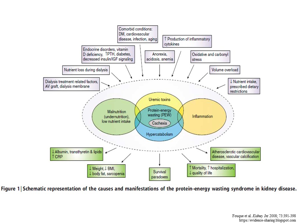
- In order to increase overall survival and compress morbidity, a multi-modal intervention approach is needed, which should target the discussed factors involved in cachexia (Figure 1).
- Such a multi-modal intervention approach, encompassing exercise training and improvement of energy balance and nutrient availability, is currently feasible as supported by recent statements and meta-analyses, possibly improved in the near future by targeted pharmacological interventions and cognitive behavioural therapy to sensitize patients to anabolic stimuli


Further reading:

Wasting in chronic kidney disease

INTRODUCTION
- Wasting is prevalent among patients with chronic kidney disease (CKD)
- About 18% to 75% of adults with end-stage renal disease (ESRD) undergoing maintenance dialysis showed some evidence of wasting
- Although inadequate nutrition may contribute to wasting or cachexia, other factors including systemic inflammation, perturbations of appetite-controlling hormones from reduced renal clearance, aberrant neuropeptide signaling, insulin and insulin-like growth factor resistance, and metabolic acidosis, may be important in the pathogenesis of CKD-associated wasting
- The wasting/cachexia syndrome in CKD patients consists of anorexia, increased energy expenditure, decreased protein stores characterized by a low serum albumin, and loss of body weight and loss of muscle mass
- Importantly, the individual components of this syndrome all represent risk factors for mortality in patients with CKD, which is 100–200 times higher than the general population
- The wasting/cachexia syndrome should be distinguished from malnutrition: 

DIAGNOSTIC CRITERIA OF CACHEXIA/PROTEIN-ENERGY WASTING IN CKD 
- Cachexia was defined as “a complex metabolic syndrome associated with underlying illness and characterized by loss of muscle, with or without loss of fat” (Evans et al., 2008)
- In the context of CKD, the term protein–energy wasting (PEW) has been proposed by The International Society of Renal Nutrition and Metabolism (ISRNM) to describe a “state of decreased body stores of protein and energy fuels (body protein and fat masses)”
- The ISRNM suggested that the term cachexia be reserved for only the most severe forms of PEW. However, there is no obvious distinction between PEW and cachexia from a pathophysiology standpoint
- Limiting the term cachexia to the extreme forms of PEW could be considered too restrictive. The term pre-cachexia has been proposed to include the milder forms of wasting in cancer patients (Fearon et al., 2011)

- Cachexia Definition (Evans et al., 2008)

- Protein-energy wasting in CKD (by ISRNM, Fouque et al., 2008)

DIAGNOSTIC CRITERIA OF CACHEXIA/PROTEIN-ENERGY WASTING IN CKD (cont’)
Anthropometric Indicators
1) BMI
- Although BMI gives little information about body composition, BMI is a useful means of assessing PEW.
- BMI is strongly correlated with LBM at the low end of the BMI spectrum, and low BMI is a consistent predictor of mortality in both adults and children on maintenance dialysis.
- However, BMI is not a very precise parameter of nutritional status in patients in whom gross imbalances in fluid homeostasis are commonly observed, such as in patients with ESRD, CHF, and liver disease.
- Furthermore, in patients with significant muscle wasting with relatively well-preserved fat mass, small changes in BMI may well be obscured by imbalances in fluid homeostasis

2) SGA
- Stenvinkel et al. analyzed 268 patients with ESRD according to their BMI and SGA,
- They found that 38% of their patients in the low BMI group had a normal SGA, whereas 45% of the patients in the normal BMI group and 17% in the high BMI group were considered to have PEW by SGA
- Low BMI has no impact on cardiovascular mortality whereas an SGA 2 was associated with a marked increase in cardiovascular mortality

3) Rate of weight loss
- Unintentional weight loss or reduction in weight of 5% or more over 3 months, or 10% or more over 6 months are suggested as indicators of cachexia/PEW, independent of absolute BMI

4) Growth Failure in Children
- Linear growth failure in children with CKD was highlighted as central to the diagnosis of cachexia, and has been associated with a greater mortality risk in children on maintenance dialysis
- However, the etiology of growth retardation in CKD is multifactorial, including other factors such as delayed sexual maturation, bone disease, acidosis, and growth hormone/insulin growth factor resistance.
- Growth failure may emerge as a necessary, but insufficient criterion, for PEW in children with CKD

5) Muscle mass
- Reduced muscle mass appears to be the most valid criterion for the presence of PEW in CKD, and is also emphasized in the diagnostic criteria for cachexia.
- Mid-arm circumference has been shown to correlate with quality of life and survival in adult patients on maintenance hemodialysis (HD).
- Dual X-ray absorptiometry, near-infrared interactance, and bioelectrical impedance have been used in investigations of ESRD patients [30] but these techniques have limitations in
ESRD and are not currently accepted as clinically useful tools.
- Indirect measures, such as creatinine appearance (estimated by quantification of creatinine in a 24-h urine collection and in the collected spent dialysate) have been proposed as an index of muscle mass in patients with CKD and ESRD

6) Fat mass
- Body fat mass lower than 10% of body weight is considered an additional criterion for PEW in adults with CKD due to the known association between total body fat below 10% and increased mortality risk in adult maintenance dialysis patients
- A more recent study showed that higher fat mass in dialysis patients might actually be protective in survival predictability
- Nevertheless, abdominal fat deposition was shown to be linked to inflammation and PEW, resulting in an increased mortality risk in maintenance HD patients

Biochemical Indicators
1) Albumin
- Low serum albumin is a consistent predictor of mortality in both adult and pediatirc ESRD patients.
- A low serum albumin concentration is by far the strongest predictor of mortality and poor outcomes in adult ESRD patients on maintenance dialysis when compared to any other risk factors, including traditional risk factors (hypertension, smoking, hypercholesterolemia, diabetes, and obesity) and nonconventional ones (anemia measures, oxidative stress, minerals and bone surrogates, dialysis treatment and technique)
- Dialysis patients with baseline serum albumin of even 0.2 g/dL higher or lower than other dialysis patients with similar demographic and comorbidity constellations have significantly lower or higher death risk, respectively.
- The albumin–death association is highly incremental and linear, and the mortality–predictability of serum albumin below 4.0 g/dL has virtually no cutoff level, below which the association with death would cease or reverse

2) Prealbumin
- Low serum prealbumin (e.g., <30 mg/dL) is another indicator of PEW and a strong predictor of outcomes in maintenance dialysis patients
- Even though baseline serum prealbumin may not be superior to albumin in predicting mortality in maintenance HD patients, prealbumin concentrations <20 mg/dL are associated with death risk even in normoalbuminemic patients, and a fall in serum prealbumin over 6 months is independently associated with increased death risk
- Dialysis patients with high serum prealbumin have lower proportion of body fat as well as higher proportion of muscle mass, which suggest that normal serum prealbumin is associated with reversal of the abnormal body composition in cachexia
- Dialysis patients with a baseline serum prealbumin between 20 and 40 mg/dl, a drop of 10 mg/ dl was associated with 37% increase in death risk independent of baseline markers of malnutrition inflammation score (MIS), serum albumin, and inflammatory markers

Food & Nutrition Indicator (Anorexia)
- Both subjectively reported anorexia as well as measured low protein or energy intake has been associated with increased mortality in adult ESRD patients
- Anorexia is prevalent, in 30–40% in adult maintenance HD patients, and is associated with higher concentrations of pro-inflammatory cytokines and higher levels of erythropoietin hypo-responsiveness as well as poor clinical outcome, including a fourfold increase in mortality, greater hospitalization rates, and poor quality of life.
- As male ESRD patients seem to be more prone to inflammation-associated anorexia than female patients, sex hormones may play an important role in this context.
-Anorexia is prevalent in children with fairly mild CKD can be the primary reason for growth failure
- Poor growth due to inadequate intake has been observed in children with glomerular filtration rate as high as 70 ml/min/1.73m2
- Growth of children with CKD is compromised when energy intake fall below 80% of recommended daily allowance.

Other indicators
- Serum transferrin, choleteral, inflammatory markers such as CRP, proinflammatory cytokine such as IL-6.

PATHOPHYSIOLOGY OF CACHEXIA/PEW SYNDROME IN ADVANCED CKD
- The pathophysiology of cachexia/PEW syndrome is CKD is multifactorial. An overview is summarized in Figure below. Please refer to the original paper for detailed discussion on Anorexia, Increased energy expenditure, Inflammation, Insulin resistance and Vitamin D deficiency

THERAPEUTIC STRATEGIES FOR PREVENTION AND/OR TREATMENT OF CACHEXIA/PEW IN CKD
1)  Nutritional supplementation
- There is evidence that nutritional therapy will improve PEW in adult ESRD patients, such as the use of oral nutritional supplement (ONS), Intradialytic parenteral nutrition (IDPN) --> improved serum albumin and/or prealbumin level.
- Growth of children with CKD is compromised when energy intake fall below 80% of recommended daily allowance (RDA). Increasing energy intake to 100% (but not more than 100%) of the RDA can increase weight gain and stabilize growth rates

2) Exercise & Physical Activity
- While that is evidence that patients with ESRD can improve skeletal muscle quality by exercise, longer training durations or more sensitive analysis techniques are required before this regimen can be recommended as therapy for cachexia/PEW in CKD.
- There is no data on whether exercises capacity tests can predict outcome in ESRD.

3) Appetite stimulant
- Megestrol acetate is a synthetic derivative of progesterone. Megestrol acetate may induce appetite via stimulation of hypothalamic neuropeptide Y, modulation of calcium channels in hypothalamic appetite centers or inhibition of inflammatory cytokines such as IL-1, IL-6, and TNF
-  In the only double-blinded, crossover study of 24 maintenance hemodialysis patients with anorexia, no significant increase in albumin or LBM was observed. A large number of side-effects were reported, including headaches, dizziness, confusion, diarrhea, hyperglycemia, thrombo-embolism, uterine bleeding, peripheral edema, hypertension, and adrenal insufficiency
- Thus, the current experience in ESRD patients does not support the use of megestrol acetate in clinical practice.

4) Correction of Acidosis
- There is evidence that acidosis can induce muscle protein catabolism and it could therefore be an important factor contributing to loss of muscle protein in these conditions
- Acidosis is associated with negative nitrogen balance and degradation of branched-chain amino acids and protein
- There are, however, few treatments available for correcting metabolic acidosis apart from alkali supplements such as NaHCO3 which, in CKD patients, carry the risk of sodium loading and fluid overload
- Despite the risk, a recent study using NaHCO3 supplementation in patients with (predialyss) CKD actually led to a slower decline in their renal function as well as improvement in their nutritional status (dietary protein and calorie intake increased, accompanied by improvements in serum albumin and LBM as assessed by mid-arm muscle circumference)

5) Growth Hormone
- Acquired resistance to the anabolic actions of growth hormone (GH) is a potential cause of the increased net protein catabolism and wasting in patients with advanced CKD.
- Studies showed that pharmacologic doses of recombinant human growth hormone (rhGH) improves whole body protein homeostasis in chronic HD patients in the short-term.
- More studies are needed to evaluate its long-term effect of rhGH on outcomes in patients with advanced CKD

6) Ghrelin agonists
- The salutary effects of ghrelin on food intake and meal appreciation suggest that it could be an effective treatment for anorexic ESRD patients
- Tolerance in appetite-regulating centres and/or other factors may override the long-term appetite-stimulating effects of ghrelin
- Ghrelin infusion acutely induces lipolysis and insulin resistance independently of GH and cortisol, thus it will be important to follow subjects for the risk of diabetes while on long-term ghrelin treatment
- A major limitation of treatment based on natural hormones is the need for parenteral administration, because of the large size of the molecule. The long-term therapeutic potential of GHS-R agonists will likely rest with orally bioavailable compounds
- Despite reports of the short- and intermediate-term success of ghrelin administration in treating anorexia and cachexia in ESRD patients, we must await results of studies on its long-term efficacy

7) Leptin and melanocortin signalling modulation
- Studies in mice shows promising results, human studies are awaited.

8) Ubiquitin-proteasome inhibitors
- Cachexia/PEW in CKD is characterized by protein catabolism. Protein synthesis is unchanged while protein degradation is increased in CKD. The daily rate of protein turnover in cells is so high that even a small increase in protein degradation will cause marked protein depletion over time.
- The mechanism of increased protein degradation in CKD is through the activation of the UPS.
- Complications of CKD, including acidosis, insulin resistance, inflammation, and increased glucocorticoid and angiotensin II production, all activate the UPS to degrade muscle protein
- Recognition of the role of the UPS in the pathogenesis of cachexia/wasting has led to the therapeutic use of bortezomib—a proteasome inhibitor—in cancer patients.
- Inhibition of the proteasome will block activation of NF-[kappa]B, which is a common final pathway for signal transduction of many cytokines, thought to be a central mechanism of cachexia/wasting in many chronic disease states including CKD

9) Dose & frequency of dialysis
- A recent randomized controlled trial failed to confirm the beneficial effects of daily HD on nutritional status in maintenance HD patients, as measured by serum albumin

CONCLUSION
- Many questions remain about the description, classification, and treatment of PEW or cachexia in children and adults with CKD.
- Whether nutritional supplementation can improve nutritional status and hence morbidity and mortality in ESRD patients remains to be tested with appropriately designed RCTs
- Most of the information on the novel strategies is currently at the experimental level and awaits confirmation by RCTs in patients with CKD-associated cachexia/PEW syndrome.


Further Reading:

Cardiac cachexia: A systematic overview

INTRODUCTION
- The incidence of CHF in the European and the United States is about 0.1-0.5% per ear; while the prevalence of CHF is about 0.3-2.4% (~5 million people) in the United States.
- About half of the patients with chronic heart failure (CHF) die within 4 years of diagnosis
- In unselected patients with CHF, mortality rates were as high as 50% in the cachectic subset compared to 17% in the non-cachectic subset at 18 months of follow-up.
- Cachexia is not only associated with poor outcomes, but also with an unfavourable response to drug treatment and poor quality of life

DEFINITION OF CARDIAC CACHEXIA
- Cardiac cachexia as a clinical entity is acknowledged as a complex syndrome, which is associated with poor outcomes.
- No single reason for cachexia exists
- Patients usually experience progressive weight loss with body composition alterations and disturbed homeostasis of several body systems.
- There is evidence for activation of neuroendocrine and inflammatory systems, increased lipolysis, muscle wasting, lack of appetite, and malabsorption whilst the importance of individual pathways and the exact interplay remain unknown.
-The authors proposed definition: non-oedematous weight loss of >6% of total body weight over a period of 6 or more months (At this stage it is important to note that the severity of cardiac cachexia may not always correlate with classical criteria of disease severity as New York Heart Association [NYHA] functional class, left ventricular ejection fraction, or exercise duration; Cardiac cachexia even may not be associated with morphological cardiac changes as seen by magnetic resonance imaging or echocardiography)
- It might be necessary to add some laboratory, clinical and functional parameters to be able to identify cachexia and body wasting in an early phase.
- Ideally, patients at risk for the development of cachexia should be identified as early as possible, however, no effective treatment of manifest cachexia is available yet.
- The definition by Evans 2008 may also be relevant.

Cautious with terms such as “cachexia”, “anorexia”, “sarcopenia”, “malnutrition” and “hypercatabolism” – they are not the same!
- Sarcopenia: age associated “normal” muscle wasting, may not result in significant weight changes, because loss of muscle and increases in fat mass are frequently balanced.
- Malnutrition and Anorexia: are associated with predominantly loss of fat mass rather than muscle tissue; are reversible with adequate food intake, but not cachexia
- Hypercatabolism cannot be evaluated during clinical examination and neglects the other side of the coin, the anabolic processes

PATHOPHYSIOLOGICAL ASPECTS OF CARDIAC CACHEXIA
1) Immune Activation
- One key aspect of CHF and cardiac cachexia like many other forms of cachexia is inflammatory immune activation
- Activation of the pro-inflammatory mediator tumour necrosis factor-α (TNFα) is the final common pathway that links all forms of cachexia.
- One of the most important signal transducers of many inflammatory stimuli is nuclear factor-κB (NF-κB).
- Interestingly, over-activity of the NF-κB system has been shown to occur in patients with CHF

2) Regulation of Feeding
- Feeding is a key component of a satiety-hunger homeostaticmodel (Fig.1).
- Although a simple but vital daily process, it is influenced by many pathways and/or mechanisms, which are still not completely understood (Table 2)
- The hypothalamus has been identified as the central regulating site of appetite (Fig. 2).
- Two areas can be differentiated: a lateral “feeding area” and a medial “satiety centre”
- Excessive fluctuations in feeding cause weight and body composition changes which may develop into medical conditions or disease
- Cachexia of chronic illnesses shares several nutritional features.
- Especially patients with cardiac cachexia experience appetite problems and alterations in food intake, malabsorption of nutrients, metabolic disturbances, and finally an anabolic/catabolic imbalance
- Malnourishment due to lack of appetite can occur in a variety of chronic diseases and as many as half of the patients can be affected.
- Disturbances in energy expenditure can contribute to anorexia-mediated body wasting and eventually to cachexia.
- Therefore, it is a key element in treating chronic illnesses that such patients receive adequate nutrition in order to prevent further development of the disease, to avoid potential side effects of treatment, and to recover from a state of deterioration.

3) Mechanisms of wasting in different body compartments
a)  Skeletal muscle is lost due to an imbalance of protein synthesis and proteolysis
- Typically proinflammatory cytokines induce proteolytic systems, while simultaneously reducing the anabolic IGF-1 signalling.
- Additionally myostatin, a negative regulator of muscle mass, is considered to be a key player, as it has been reported as up-regulated in HIV cachexia and sarcopenia

b) Later in the course of the disease, wasting of bone and fat mass is found
- Fat wasting can be induced by numerous agents and the rate limiting step is the activity of the enzyme hormone sensitivity lipase that can be activated by several receptors including β-adrenergic and natriuretic peptides receptors.
- TNFα, which is also over-expressed in both CHF and cardiac cachexia, plays a major role in fat cell lipolysis and inhibiting insulin signalling. The latter is an important aspect in the development of insulin resistance.
- It is tempting to speculate that increased levels of catecholamines and possibly natriuretic peptides may be responsible for the loss of adipose tissue that has been observed in such patients
- The bone density in patients with cardiac cachexia is lower, and lower calcium and vitamin D levels have been reported, although no mechanisms have been identified so far

- These alterations in body composition have their reasons in profound metabolic perturbations, which are initially meant to isolate and neutralize the insult that caused the heart to fail.
- However, at a later stage, they contribute to the development and the progression of cardiac cachexia.

THERAPEUTIC APPROACHES TO CARDIAC CACHEXIA
- Currently there are no approved therapies to treat of weight loss as such in cardiac cachexia

1) Prevention of Weight Loss
a) ACE Inhibitors & Beta-blockers
- Angiotensin Converting Enzyme (ACE) inhibitors and beta-blockers have both clinically shown their potential to delay and possibly prevent the onset of cardiac cachexia.

2) Nutrition
a) Feeding
- Although feeding alone, the reversal of the clinically observed anorexia, does not reverse cachexia, nutrition itself still seems to be a major factor in treatment strategies
- Often patients have deficiencies in micronutrients such as vitamins and a supplementation of branched-chain amino acids has shown to be beneficial
- An overall induction of food intake to battle anorexia in these patients is considered supportive.

b) Nutritional Consideration
- However, the daily sodium intake should be restricted to 2 g in all patients with advanced CHF or cardiac cachexia
- Prolonged periods of fasting are potentially harmful, and cachectic patients should be advised to eat small, frequent meals
- Fluid intake should be restricted to 1.5–2.0 l/day, especially in patients with severe symptoms or those requiring high doses of diuretics
- At least one study suggests that multiple micronutrient supplementation is potentially beneficial. Such supplements should contain anti-oxidant supplements and B-group vitamins
- Avoid food and lifestyle factors that trigger the acute phase response such as an excess of carbohydrates or saturated fats, alcohol, and smoking
- Food that counteracts inflammatory responses, can be recommended. This includes fish oil supplements, olives, walnuts, flaxseed oil, any fruits or vegetables, garlic, ginger, turmeric, sunflower seeds, eggs, herring, or nuts
- Enteral nutrition should always be given preference over parenteral nutrition, however, if the latter cannot be avoided, the general guidelines can be followed: 35 kcal per kg of bodyweight per day,1.2 g of protein per kg per day, and a 70:30 glucose:lipid ratio for the non-protein energy (ZYL note: I recommend to start slow while monitor for refeeding syndrome; increase calorie intake slowly as tolerated and prevent overfeeding)

3) Pharmacotherapy

I) Appetite Stimulants
a) Megestrol Acetate
- Megestrol acetate has been shown to stop weight loss in hormone responsive cancer and has recently been approved for AIDS cachexia in the United States.
- The closely related compound, medroxyprogesterone acetate, has similar effects, but a trend towards peripheral oedema was observed. Cannabinoids

b) Cannabinoids
- Cannabinoids are known inducers of food intake, but have clinically shown contradictory results.
- While positive effects on appetite were seen in patients with AIDS and cancer, there was no concomitant weight gain.

II) Anabolic agent
a) Anabolic Steroids
- The use of anabolic steroids in cachexia patients is limited to chronic obstructive pulmonary disease and AIDS, where positive effects on weight have been observed
- In CHF, there was an improved cardiac function, but no effect on weight

b) Beta-adrenergic agonist
- Beta-adrenergic, especially β2-adrenergic agonists are also known for their anabolic properties and have shown some beneficial effects in degenerative muscle diseases.
- Although short-lived, positive cardiac effects have been reported in CHF, but there was no improvement in the functional capacity of the patients

III) Anti-inflammatory strategies
- a) Neutralizing antibodies like eternacept and infliximab, which showed a lack of efficiency in CHF,
- b) Statins, which show beneficial pleiotropic effects like the reduction of pro-inflammatory cytokines in some settings and
- c) Thalidomide, which potently reduces TNFα

IV) Inhibition of major proteolysis pathway
- The use of proteasome inhibitors to inhibit major proteolysis pathway, the ubiquitin-proteasome pathway is in its early stages and has so far only been tested in cancer, where weight gain has been reported.
- While pentoxifylline reduced the activity of muscle proteolytic systems in a rat model of cancer cachexia, it failed to improve weight in patients with cancer cachexia.

CONCLUSION
- The pathophysiology of cardiac cachexia is exceedingly complex, and we still do not understand when and how CHF progresses into this syndrome
- Pro-inflammatory cytokines and especially TNFα certainly play an important part. However, therapies that targeted specific single cytokines have largely failed, and it appears that broader approaches are required.
- We are currently not able to interfere with appetite regulation in a promising way, although initial steps have been undertaken.
- Nutritional recommendations for cardiac cachexia remain speculative, and no large-scale randomized, controlled trials have been performed